Kanaa Fertility Centre is one of the top-rated clinics in Chennai providing Sonohysterography. Our team of experts helps patients interested in knowing what is SSG and it benefits before the actual procedure. Book Now!
Sonohysterography is a procedure to look at the inside of the uterus. It’s a safe, painless test that uses sound waves and a computer to create images. It does not use radiation. Sonohysterography does not use ionizing radiation, has no known harmful effects, and provides a clear picture of soft tissues that don’t show up well on x-rays.
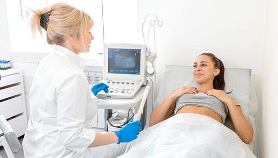
Procedure: A thin, flexible tube (catheter) is inserted into the cervix. Saline is sent through the catheter into the uterus. At the same time, the transducer sends high-frequency sound waves through the gel and into the body. The echoes from these sound waves create a real-time image of the inside of the uterus. This shows the structure of the uterus. The saline fluid helps the ultrasound form an image with sharper detail. This information helps to diagnose a number of different health conditions
The flow of fluid through the tubes into the pelvis when saline is injected can be visualised on the ultrasound and hence is used to determine tubal patency

SSG (Saline Sonosalpingography) is an ultrasound-based test used to evaluate the uterus and fallopian tubes. During the procedure, sterile saline or contrast fluid is introduced into the uterus while ultrasound imaging checks whether the fallopian tubes are open. SSG is commonly recommended during fertility evaluation before treatments such as IUI or IVF.
When couples face difficulty conceiving, doctors often begin with diagnostic tests that evaluate reproductive health. One important test used in fertility evaluation is the SSG test, also known as Saline Sonosalpingography.
SSG helps fertility specialists examine the uterus and determine whether the fallopian tubes are open. Because the fallopian tubes play a crucial role in natural conception, identifying blockages or abnormalities early helps guide the most appropriate fertility treatment.
Understanding how the SSG test works and what it evaluates can help patients feel more comfortable during the diagnostic process.
SSG (Saline Sonosalpingography) is a diagnostic fertility test performed using ultrasound imaging. It allows doctors to examine:
During the procedure, sterile saline or contrast fluid is gently introduced into the uterus through a thin catheter. Ultrasound imaging tracks how the fluid flows through the uterus and fallopian tubes.
If the tubes are open, the fluid moves freely into the abdominal cavity, indicating normal tubal function.
The SSG test helps identify possible causes of infertility by evaluating the uterus and fallopian tubes.
Doctors may recommend an SSG test to:
SSG is often performed as part of a complete fertility assessment.
Doctors may suggest an SSG test when:
The test is typically done after menstruation and before ovulation.
The SSG test is usually scheduled in the early part of the menstrual cycle to ensure the uterus is clearly visible and pregnancy is not present.
Patients may be advised to:
During the test:
If the fluid flows freely through the tubes, it suggests that the tubes are open.
The entire SSG test usually takes 15–20 minutes and is performed as an outpatient procedure.
When both fallopian tubes are open and the uterus appears normal, doctors may recommend natural conception attempts or treatments depending on other fertility factors.
If fluid does not pass through the tubes, it may indicate a blockage. This condition may prevent sperm and egg from meeting naturally.
Doctors may recommend further evaluation or assisted reproductive treatments.
SSG may reveal abnormalities in the uterine cavity such as:
Further evaluation with Hysteroscopy may be recommended to diagnose and treat these issues.
Both SSG and HSG evaluate fallopian tubes, but they use different imaging methods.
Test | Imaging Method | Purpose |
HSG | X-ray | Detect tubal blockage |
SSG | Ultrasound | Evaluate uterine cavity and tubes |
Doctors choose the test based on medical history and diagnostic needs.
Most women experience mild discomfort similar to menstrual cramps during the procedure. The discomfort is usually temporary and subsides shortly after the test.
Many patients find SSG more comfortable than X-ray based tests.
SSG is generally considered a safe procedure. Possible side effects may include:
Doctors provide instructions to minimize risks and ensure patient safety.
In some cases, the saline or contrast fluid used during the test may help clear minor debris from the fallopian tubes. While this is not guaranteed, some couples conceive naturally after the procedure.
However, the primary purpose of the SSG test is diagnosis, not treatment.
The SIS procedure is conducted in a fertility clinic under the guidance of an expert:
Although routine ultrasound of the uterus is a helpful screening test, it may not delineate the lining of the uterus or minor abnormalities. A Sonohysterogram (SIS test) or saline infusion sonogram employs saline infusion to fill up the uterine cavity. This yields a clearer, more precise view of the uterus so that embryologists and gynecologists can pick up on fine uterine abnormalities that a standard uterus ultrasound might not. Uterus sonography with saline renders the best visualization for fertility testing.
Following a saline infusion sonogram, the pictures are examined closely by your fertility expert. The uterus ultrasound pictures formed throughout the SIS test can determine:
Clear imaging of SIS findings assists in planning fertility treatment, directing interventions, or determining the causes of infertility.
A Sonohysterogram, also called a saline infusion sonogram, is a safe procedure that uses saline infusion and uterus ultrasound to create detailed images of the uterine cavity. It helps detect fibroids, polyps, scarring, or abnormal uterine shapes.
Sonohysterography is prescribed to find the underlying cause of many problems, including abnormal uterine bleeding, infertility, and repeated miscarriage.
In a patient with regular menstrual cycles, sonohysterography should, in most cases, be performed by day 10 of the menstrual cycle. In cases of intermittent or prolonged abnormal uterine bleeding, treatment with a short course of progestin may be considered to enable timing of the procedure.
Other tests may be used to diagnose problems of the uterus:
Sonohysterography has some benefits over other tests used to get information about the uterus. Other tests include:
· Hysterosalpingography. This is a type of X-ray that uses radiation and a fluid that is a radioactive dye.
· Hysteroscopy. This is a surgical procedure that needs to be done with anaesthesia. But this procedure also helps to treat certain problems when it is diagnosed.
· MRI. This is an imaging test done with large magnets and a computer. An MRI may not give a clear picture of the inside of the uterus.
Sonohysterography is limited in the assessment of the patency, or openness, of the fallopian tubes because of their size and structure. In cases your doctor suspects an abnormality of the fallopian tubes, they may recommend Hysterosalpingography or diagnostic hystero lap for further evaluation.
Most women are able to go home right away and are back to their normal activities that day. Some of the following symptoms may occur after the test:
· Cramping
· Spotting or light bleeding
· Watery discharge
Most patients experience mild cramping or discomfort during the saline infusion sonogram, but it is generally well-tolerated and considered a minimally invasive test.
The SIS test can identify:
It is typically done after menstruation but before ovulation (days 6–12 of the cycle), when the endometrial lining is thin for optimal visualization.
Yes, the flow of saline infusion through the fallopian tubes can be observed during the procedure, helping evaluate tubal health for fertility treatments.
The procedure is very safe. Mild cramping or spotting may occur, but SIS has no radiation and no known harmful effects.
It is often recommended during fertility evaluation to assess tubal health before treatments like IUI or IVF.
The procedure usually takes around 15–20 minutes.
Yes, many couples can resume attempts to conceive shortly after the procedure.
Both tests are safe when performed under medical supervision. The choice depends on clinical needs.